


Symptoms of MCAS
MCAS symptoms arise from inappropriate mast cell activation. Activated mast cells release inflammatory mediators, including histamine, prostaglandins, leukotrienes and cytokines. The pattern of mast cell mediator release is influenced by what triggers activation. This in turn influences the type of symptoms patients experience
Symptoms typically wax and wane, with severity fluctuating over time. Exacerbations may follow identifiable triggers such as infection, surgery, hormonal changes, medical procedures, or significant physical or psychological stress. Most patients experience multiple co-occurring symptoms across several body systems, and the cumulative burden can be substantial, with marked impact on daily functioning and quality of life.
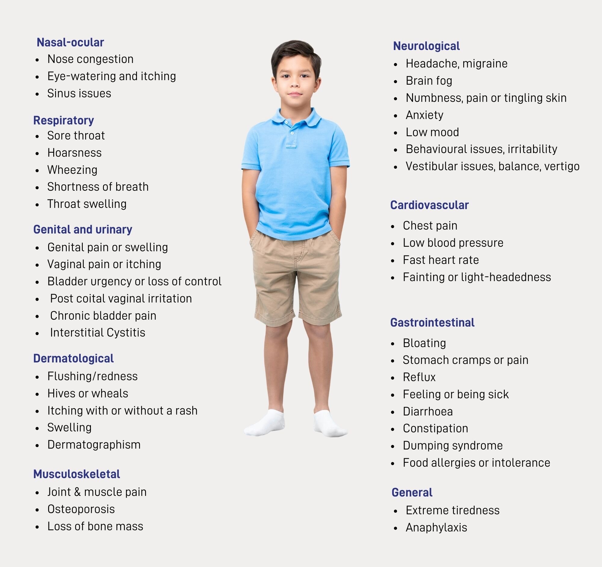
Patients’ accounts of their symptoms and patterns over time can provide important and reliable information alongside clinical clues.
The systemic nature of MCAS means it can be missed if only one issue or symptom is considered at a time. A correct diagnosis is more likely when symptoms are considered collectively, as part of a broader clinical picture.
MCAS causes symptoms in multiple organ systems.
For further information about specific systems, please use the links above.
Major human mast cell–derived mediators
| Class | Mediators | Physiological effects |
|---|---|---|
Preformed mediators | Histamine, serotonin, heparin, neutral proteases (tryptase and chymase, carboxypeptidase, cathepsin G), major basic protein, acid hydrolases, peroxidase, phospholipases | Vasodilation, vasoconstriction, angiogenesis, mitogenesis, pain, protein processing/degradation, lipid/proteoglycan hydrolysis, arachidonic acid generation, tissue damage and repair, inflammation |
Lipid mediators | LTB4, LTC4, PGE2, PGD2, PAF | Leukocyte chemotaxis, vasoconstriction, bronchoconstriction, platelet activation, vasodilation |
| Cytokines | TNF-α, TGF-β, IFN-α, IFN-β, IL-1α, IL-1β, IL-5, IL-6, IL-13, IL-16, IL-18 | Inflammation, leukocyte migration/proliferation |
| Chemokines | IL-8 (CXCL8), I-309 (CCL1), MCP-1 (CCL2), MIP-1αS (CCL3), MIP1β (CCL4), MCP-3 (CCL7), RANTES (CCL5), eotaxin (CCL11), MCAF (MCP-1) | Chemoattraction and tissue infiltration of leukocytes |
| Growth factors | SCF, M-CSF, GM-CSF, bFGF, VEGF, NGF, PDGF | Growth of various cell types, vasodilation, neovascularization, angiogenesis |
* The mediators in the table are examples only. In addition, many mediators are identified in human mast cell lines or primary cultures of human mast cells and may not be produced in vivo.
There are 2 types of mast cells found in the body, connective tissue and mucosal mast cells.
Mucosal mast cells are located mostly in the respiratory tract and gastrointestinal tract but can also be found in the blood and connective tissue.
Mast cells present IgE antibodies on their surface which bind to foreign bodies (usually allergens) causing antibody-induce degranulation, this releases mediators such as histamine.
Mastocytosis is the pathologic clinical presentation can occur from clonal mast cell gain-of-function mutations.
The most common mutation is KIT, found in transmembrane receptor tyrosine kinase.
KIT mutations cause the accumulation of mast cells in tissues, interfering with surrounding tissues and causing systemic and local interference:
- In patients presenting with Mastocytosis, dense infiltration of mast cells aggregates in bone marrow and organs. Secondary MCAS in mast cell hypersensitivity and overstimulation of mediators occurs in reaction to an external factor
- The mast cell is designed to be the first response to infection and injury, it recruits secondary immune cells and releases numerous proteins, hormones and chemicals to support the innate and active immune systems. In patients with MCAS, these reactions are excessive and damaging to the surrounding tissue, as well as systemically.
Typical symptoms include two or more organ systems with a range of systems.
Systemic and general symptoms of mast cell activation syndrome
Skin | Muscles | Bone | Urinary | Genitals | Brain | Heart | Face | General | Respiratory | Gastrointestinal |
|---|---|---|---|---|---|---|---|---|---|---|
Hives Swelling Rashes Flushing Itching | Muscle pain Muscle fatigue Numbness Tingling | Joint pain Brittle bones | Pain when urinating Bladder urgency Loss of control of bladder | Pain Swelling Discharge Itching | Headaches Anger Anxiety Brain fog | Pain in the chest Lightheadedness Low blood pressure Tachycardia (fast heart rate) | Congestion Itchy eyes Watery eyes Swelling | Fatigue Redness Feeling unwell | Anaphylaxis* Airway constriction Increased mucous Coughing Increased susceptibility to infection Asthma-like symptoms Allergic asthma | Diarrhoea Constipation Cramping Reflux Dumping syndrome** Abdominal cramping. |
Idiopathic anaphylaxis is a MCAS symptom where the patient meets the diagnostic criteria through anaphylaxis. These severe cases may present with difficulty breathing, swelling, hives and other immediate respiratory responses.
* Noticed swelling of the tongue, lips and throat. This may be accompanied with difficulty breathing, noticeable wheezing, coughing and feeling faint. These symptoms are serious and require immediate medical attention and the use of adrenaline.
** Symptoms related to gastric dumping include weakness, vertigo, tachycardia (rapid heart rate) and abdominal cramping. This rapid emptying of the stomach can be triggered by food that is low in fibre, high in sugars or simple carbohydrates.
References
- Robey RC, Wilcock A, Bonin H, Beaman G, Myers B, Grattan C, et al. Hereditary alpha-tryptasemia: uk prevalence and variability in disease expression. J Allergy Clin Immunol Pract. 2020;8(10):3549–56.
- Akin C. Mast cell activation syndromes. Journal of Allergy and Clinical Immunology [Internet]. 2017 Aug [cited 2023 Jul 20];140(2):349–55. Available from: https://linkinghub.elsevier.com/retrieve/pii/S0091674917310254
- Fellinger C, Hemmer W, Wöhrl S, Sesztak-Greinecker G, Jarisch R, Wantke F. Clinical characteristics and risk profile of patients with elevated baseline serum tryptase. Allergologia et Immunopathologia [Internet]. 2014 Nov [cited 2023 Jul 20];42(6):544–52. Available from: https://linkinghub.elsevier.com/retrieve/pii/S0301054614001220
- Butterfield J, Weiler CR. The utility of measuring urinary metabolites of mast cell mediators in systemic mastocytosis and mast cell activation syndrome. The Journal of Allergy and Clinical Immunology: In Practice [Internet]. 2020 Sep [cited 2023 Jul 20];8(8):2533–41. Available from: https://linkinghub.elsevier.com/retrieve/pii/S2213219820301811
- BSACI. 2021. Mast Cell Disorders Webinar - BSACI. [online] Available at: https://www.bsaci.org/education-and-events/webinars/mast-cell-disorders-webinar. [Accessed 17 September 2021].
- Utility of urinary prostaglandins (DM, D2, F2α) & N-methyl histamine (NMH) measurements across mast cell disorders – Dr Bethan Myers, Dr Pavel Chudakou. Presented at the British Society of Haematology Virtual Annual Scientific Meeting, 25 – 28 April 2021.
- Sala-Cunill A, Cardona V, Labrador-Horrillo M, Luengo O, Esteso O, Garriga T, et al. Usefulness and limitations of sequential serum tryptase for the diagnosis of anaphylaxis in 102 patients. Int Arch Allergy Immunol [Internet]. 2013 [cited 2023 Jul 20];160(2):192–9. Available from: https://www.karger.com/Article/FullText/339749
Luskin KT, White AA, Lyons JJ. The genetic basis and clinical impact of hereditary alpha-tryptasemia. The Journal of Allergy and Clinical Immunology: In Practice [Internet]. 2021 Jun [cited 2023 Jul 20];9(6):2235–42. Available from: https://linkinghub.elsevier.com/retrieve/pii/S2213219821003068
Batiha GES, Al-kuraishy HM, Al-Gareeb AI, Welson NN. Pathophysiology of Post-COVID syndromes: a new perspective. Virology Journal [Internet]. 2022 Oct 9 [cited 2023 Jul 3];19(1):158. Available from: https://doi.org/10.1186/s12985-022-01891-2
Weinstock LB, Brook JB, Walters AS, Goris A, Afrin LB, Molderings GJ. Mast cell activation symptoms are prevalent in Long-COVID. Int J Infect Dis [Internet]. 2021 Nov [cited 2023 Jul 1];112:217–26. Available from: https://www.ncbi.nlm.nih.gov/pmc/articles/PMC8459548/
Metcalfe, Dean D. ‘Mast Cells and Mastocytosis’. Blood, vol. 112, no. 4, Aug. 2008, pp. 946–56. DOI.org (Crossref), https://doi.org/10.1182/blood-2007-11-078097.
Valent, Peter, et al. ‘Definitions, Criteria and Global Classification of Mast Cell Disorders with Special Reference to Mast Cell Activation Syndromes: A Consensus Proposal’. International Archives of Allergy and Immunology, vol. 157, no. 3, 2012, pp. 215–25. DOI.org (Crossref), https://doi.org/10.1159/000328760.
Sala-Cunill A, Cardona V, Labrador-Horrillo M, Luengo O, Esteso O, Garriga T, et al. Usefulness and limitations of sequential serum tryptase for the diagnosis of anaphylaxis in 102 patients. Int Arch Allergy Immunol [Internet]. 2013 [cited 2023 Jul 20];160(2):192–9. Available from: https://www.karger.com/Article/FullText/339749
Luskin KT, White AA, Lyons JJ. The genetic basis and clinical impact of hereditary alpha-tryptasemia. The Journal of Allergy and Clinical Immunology: In Practice [Internet]. 2021 Jun [cited 2023 Jul 20];9(6):2235–42. Available from: https://linkinghub.elsevier.com/retrieve/pii/S2213219821003068
Author: Lauren Kelly, Medical Communications and Education Lead at Mast Cell Action
Become a friend
Sign up to become a Friend of Mast Cell Action so we can keep you up to date on our progress and on how to get involved in our latest campaigns and initiatives.
Donate
Mast Cell Action relies entirely on the generosity of people like you. Please make a donation now and together we can make a difference to those affected by MCAS.